Transforming Science
When the life science research equipment you are building a home for is recognized for revolutionizing structural biology and deemed the “research method of the year” by Nature magazine — not to mention is a more than $10 million investment — the facility to host it needs to function very, very well. It helps if it looks good, too.
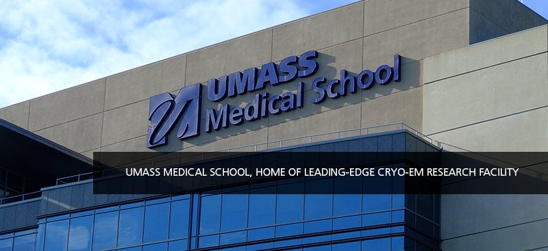
“Everybody wants it,” Chen Xu, PhD, says of the Titan Krios cryo-electron microscope (cryo-EM). Xu, who is a physicist, materials scientist and electron microscopist, has been appointed Director of UMass Medical School’s first cryo-EM microscopy facility — also the first-of-its kind in New England. This new breed of cryo-electron microscope is able to process images of particles at near-atomic level by first plunging the sample into liquid nitrogen. Opening this fall, the facility will further research for a consortium of academic and commercial partners.
Built and designed by the team of Consigli Construction Co., Inc. and healthcare architects TRO, it is not surprising that the construction of this cryo-EM facility requires a level of precision on par with the research equipment it hosts, as well as ultra-clear communication between scientists like Xu and the project team.
Crucial: Controlling the Environment
Internationally recognized for his cryo-EM expertise, Xu explains what is most crucial, “This machine is very cutting edge, it is very delicate, so we need its environment to be perfect: vibration control, the room acoustics — how quiet it is in the room — assuring a stable temperature, and that it’s protected from electromagnetic interference. All this needs to be done precisely. We are very picky.”
One of the most important new tools for drug design and development, cryo-EM is playing a critical role in identifying likely therapeutic approaches to a broad range of diseases, particularly neurological disorders like Alzheimer’s, immunological disorders and diabetes. “In the past four years, advances have made this technology very applicable — all the drug designers, all the pharmaceutical companies are rushing to it,” explains Xu.
At 4,100-square-feet, the heart of the new facility is its 2,000-square-foot microscopy suite, which hosts the facility’s two microscopes: the Titan Krios, one of the most sophisticated cryo-EMs in the world, as well as the powerful, versatile Talos-Arctica. Xu explained that the microscopes’ environment can only have a 0.1 degree change in an hour, and that the relative humidity must always be below 20 percent. To oversee these conditions, the suite is being built with round-the-clock remote monitoring systems so Xu and his team will know if there is any variation in the rooms’ environment.
Eight Layers of Protection
To build this facility to its exacting requirements for vibration, acoustical and climate control, the team is essentially building a five-roomed box, constructed with eight-layered walls, floors and ceilings, encompassing the rooms which host the microscopes as well as two observer viewing rooms. Almost two feet thick, this layered construction isolates and protects the microscopes and the immediately adjacent spaces that support their use, from noise, sound and movement, creating the stable environment that is essential to produce accurate research results.
“There’s a lot of science in this design and construction,” says TRO’s Project Architect Jacob Levine.
Consigli’s Project Manager, Mark Morrow, agrees. Describing the complexity and purpose of this layered construction, Morrow gives an overview, “If you look at an architectural cross section of the suite, you see that these eight layers include an outer layer of one inch gypsum wallboard, next there are six-inch wood studs. It is notable that we are using wood studs —rather than the more typical metal studs —which we can’t use here because of the need to eliminate vibration.
Morrow continues, “Over the wood studs is a layer of three-quarter inch plywood to which is attached a quarter-inch thickness of aluminum sheeting. These sheets then must be welded together seamlessly, with the welds ground down so the surface is completely smooth—this is essential to protect the space from electromagnetic interference. Then, over the aluminum panels we have installed one-inch-thick acoustical drywall panel, ‘Quiet Rock,’ which incorporates a damping technique called ‘constrained-layer damping,’ creating a higher ability to dampen vibrational and acoustical energy. Next come four-inch-thick ‘Sonex’ foam acoustical panels — they almost look like gym mats. Then we attached three-inch wood block anchors — a special piece custom-designed by our in-house carpentry team — to the Sonex panels to assure the secure attachment of the final layer, the four-inch-wide cooling panels.”
Taking Care, Building for Healthcare
Morrow also noted that when the high quality of welding work required for the metal panels is done within a healthcare facility, it requires special mitigation to limit smoke and smell. “While we are building the microscope facility in its ideal location from the point of view of creating a stable environment — at the basement level — making sure we created an effective strategy to ventilate any smoke and smell created by the welding, was important so it wouldn’t affect the healthcare environment.”
Morrow adds, “One of the most important aspects has been our coordination with all the trades and specialty subcontractors needed to create this special environment — not to mention working with the facilities director, Chen Xu, and our UMass user group.
A Specialized Team
“The go-to man at the center of all this is our Superintendent, Tim Backlin. Tim is our glue, he coordinates all the detail and connects with literally everyone — from responding to concerns raised by Chen, to overseeing our in-house carpentry team, to working with the specialists that the microscopes’ manufacturer recommends.” Bringing highly specialized skill to the project, these firms include the electromagnetic shielding company, Vitatech Electromagnetics — working with the team on the reduction of electron magnetic waves — as well as vibration control expert, TMC, who is helping the team take care of any disturbance in the building that would be in danger of decoupling the microscopes.
Describing the collaboration with the Consigli/TRO team, Xu noted, “They are very professional, and open to all the questions, worries from our end. We want researchers to be able to use the microscopy rooms in the most comfortable, most efficient ways possible. So it is important to consider every detail. You need to consider ‘Where will this port come into the room?‘ ‘How do we create a location for the liquid nitrogen tanks so they can be easily replaced without accidentally interfering with the microscopes?’ It is so important to understand and build this from the users’ point of view. It’s so important, if you miss the details then in the end you have a problem. Just yesterday I saw the Superintendent Tim Backlin and the team working with the acoustical isolation foam panels, they needed to cut them. They cut it so precisely. That kind of professional workmanship, I like to see it. I took pictures! This is a real detailed business, it matters very much if you are missing details for this kind of project — it can ultimately have an effect on the data.”
Xu, who intends to write a guideline for the building of future cryo-EM facilities, also emphasized how valuable it is for scientists like him to see three-dimensional design images during the design process, “Because we are not as familiar with looking at drawings, virtual views of the spaces helps users understand what is intended. It is good for the communication process.”
Data Critical
To support the work of this equipment, the facility also includes a lab for the preparation of research samples, staff offices, a large mechanical room, room for a possible third microscope, and the facility’s extensive computer resources. Xu explains, “We are talking about a lot of data that results from the cryo-EM data gathering process. To see the particle’s structure we usually need about 100,000 particle projections, the same particle showing up in all these different orientations. The critical point is your data. If the data is not good, you won’t get anywhere with your research.” With this central concern for data quality, Xu and his team will train researchers to properly prepare their samples, too.
Delivered this summer to the medical school in 19 crates, the new Titan Krios microscope will be assembled and tested over the next two months, in preparation for the facility’s opening this fall. Once assembled, the box-like form of the Titan Krios will measure close to 13 and a half feet tall — and gathering data for each sample will take four-to-five days.
This is not the microscope of high school bio lab days. The purchase of the Titan Krios system is supported by a five million dollar grant from the Massachusetts Life Science Center, while the facility’s second microscope system, the Talos-Arctica, is being paid for by the Howard Hughes Medical Institute.
With some good fortune — and continued precision — it’s exciting to imagine what the cryo-EM’s research results will offer the future.
View our full case study here.

